Confidence: 5
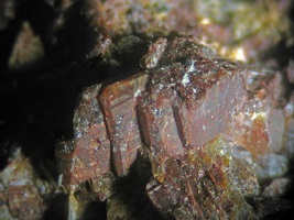
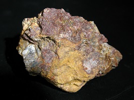
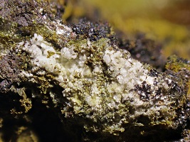
Chemistry: ThO2
Locality: Black Cap Mtn., Conway, NH
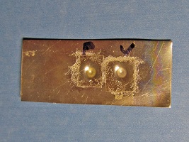
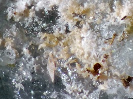
Specimen Size: Group of tabular stacked crystals, 1 cm fov. (close-up). Full specimen - 5 cm
Field Collected: Henry Berbour, obtained in trade by Gene Bearss, 1986. Gifted to Tom Mortimer
Owner: Tom Mortimer
Catalog No.: TBC
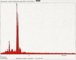
Analysis: The reddish coating appears to be on the surface of these tabular crystals... the interior, exposed in broken areas, is sort of a pale tannish-yellow, glassy. There are some black ferrocolumbite blebs on one side of the specimen. Tiny, sharp, zircon crystals are also present. The thorianite identification is by semi-quantitative XRF on an unpolished grain. ( pdf file ) - the view must be greatly expanded to see the element designations. The Iron response is likely due to surface oxide coating.
This specimen maxes out my scintillometer range at about 10 inches away.
With my Victoreen GC:
600 cpm at 5 inches, window open
1300 cpm at 3 inches, window open
300 cpm at 3 inches, window closed.
Notes: Gene had this specimen labeled as "Thorite." Thorite is a thorium silicate. No silicon was detected in the XRF spectrum, leading to the conclusion that this is thorium oxide, thorianite.
Thorianite has not been previously reported from New Hampshire. Thorite (the silicate) has been on NH mineral species lists for decades. The National Museum of Natural History has a Conway, NH thorite specimen in their collection. It was acquired in 1976 as a gift from Joe Pollick. An XRD analysis was done by Pete Dunn, (no chemical analysis was done.)

Confidence: 5
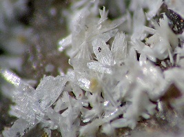
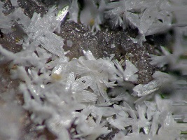
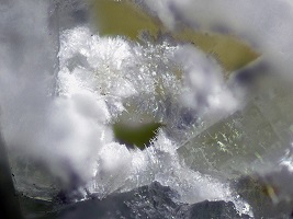
Chemistry: C5H4N4O3 · 2H2O
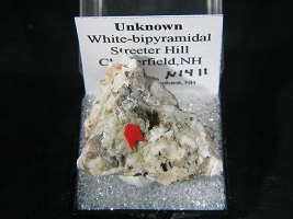
Locality: Streeter Hill Fluorite Prospect, Chesterfield, NH
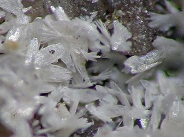
Specimen Size: Top photo, 1.5 mm field of view. Feathery crystals to 0.2 mm
Field Collected: Tom Mortimer - 2006
Catalog No.: u1411
Notes: Only one specimen of this mineral was found. Given the extreme fragility of this sample, it is (1) Fortuitous that I recognized it as interesting when I found it in the field, (2) Somewhat remarkable that I was able to get it home intact, (3) Secure it undamaged into a TN perky box, and (4) Perservere to a definitive identification.
When Kerry Day (Ottawa EDS service) looked at this, all he could see was "noise" with his instrument's limited light element detection capabilities. He suggested it was an organic. (the sample he analyzed was on carbon tape, so his result was somewhat of a "mushed mess"). These are definitely distinct feathery crystals. The substrate is quartz. These crystals are soft an friable. Some of the C on the Day analysis may have come from the mineral or from the carbon tape, or both.
The only elements that showed up on a Jan. 2015 Boston College EDS analysis were nitrogen, oxygen, and carbon. This instrument has good light element detection capability. Looking on mindat, there are a number of minerals with just N, O and C. I realize I could not eliminate Be or Li, but I think both of these are unlikely at this locality, a hydrothermal fluorite vein. Other minerals I found here were barite, fluorite, galena, and malachite. As this specimen was collected close to the surface I wondered if animal/bird dung/urine could be a contributor?
These crystals are not soluble in water or muriatic acid. They are not fluorescent.
In mid 2015, I learned of “A world-wide hunt for new carbon minerals, The Carbon Mineral Challenge", begun earlier that year. This research group arranged for a single crystal XRD of my unknown by Harvard University. I received results within two weeks of submitting my sample!
The Harvard analysis indicated the probable mineral was tinnunculite, C5H4N4O3· 2H2O
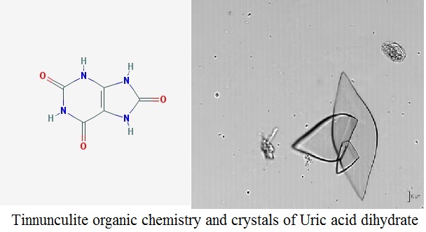
This substance has been known to science prior to the announcement of tinnunculite, (IMA approved 2015). To the biological sciences it is known as “Uric acid dihydrate”, a principal component of kidney stones. My specimen was found very close to the surface. There is a bit of moss or lichen on it. This NH tinnunculite is very likely the result of some mammal or bird urination.
There are some caveats:
* The Harvard XRD was only a partial data set.
* A question remains as to which crystal space group the Streeter Hill sample belongs to.
* A description of tinnuculite has just recently been announced, (Mineralogical Magazine, February 2016, Vol. 80(1), pg. 202).
Reference mindat web page: mindat.org link
The crystal morphology of the Streeter Hill tinnunculite is a very good match for the uric acid dihydrate crystals shown in the lower photo.
Given I found this in 2006, I had the opportunity to claim a new mineral. However, it is just as well the Russsians described it first, as I would not want “mortimerite” to be an animal piss evaporate !
History of the Tinnunculite Identification
The contact from The Carbon Mineral Challenge, Daniel Hummer of the Carnegie Institute of Science, recommended a single crystal XRD and put me in touch with Shao-Liang Zheng, Director of X-Ray Laboratory, Harvard University.
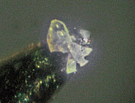
Getting a sample to Harvard presented a substantial challenge. I was able to get a tiny group of crystals onto the end of a toothpick (photo), but how to get a sample to Harvard? Following discussions with Shao-Liang, I settled on sending the crystals emmersed in oil. (See photo of my oil-dropplet carrier.) Two days later, I learned my postal mailing was successful. Shao-Liang was able to mount a crystal, collect XRD data and forward result to Daniel Hummer at Carnegie. An email from Daniel stated:
"It turns out this material corresponds to the mineral tinnunculite, which was just approved by the IMA this February (and was one of the 5 possible known C-N-O-H minerals I mentioned in a previous message). It is actually monoclinic with space group P21/c, but has a beta angle of 91.0 degrees, so it is "almost" orthorhombic. ...
It has apparently been known from a locality in Russia, the Khibiny Massif, for quite some time and has been submitted to the IMA before, but was previously rejected because it was thought that human influence contributed to the formation. a Russian group was recently successful in convincing the IMA that the occurrence was fully natural, so it was just approved as a new mineral species. So, what Tom has is the second known natural occurrence of tinnunculite."
A follow-on email exchange between Shao-Liang and Daniel Hummer expressed an uncertainty about the crystal space group of the Streeter Hill sample: whether it is "P21/c" or "Pnab".
The Carbon Mineral Challange research group had an exhibit at Tucson, 2017. Joylon Ralph snapped a photo reporting the Russian tinnuculite, reproduced here. However, the parent photo for this poster image on mindat.org (zoom view included here, crystals to 0.2 mm, copyright Uwe Kolitsch, used here with permission) indicates this specimen is from "Power station, Sportgastein, Naßfeld valley, Gastein valley, Hohe Tauern, Salzburg, Austria." I could find no photo of the Russian material, but by the standard of this Austrian sample, my New Hampshire find is not to shabby! Uwe pointed out that tinnunculite is visually similar to uricite. It has a similar chemistry as well.
It is interesting to note the IMA's initial resistance to accepting tinnunculite as a species was due to the "thought of human influence" on the mineral formation (coal mining and later coal dump fire). Given that there was no coal mining or evidence of smeltering at the small Streeter Hill Prospect, this New Hampshire example more closely fits the formal definition of a mineral. One might speculate that if heat is necessary to form this species, perhaps a brush or forrest fire occurred at this prospect some time in the past.
As a further note, per mindat.org, tinnunculite has now been reported from five different localities; one in Russia (TL), two in Austria, two in Italy, and one in Norway.
Confidence: 4
Chemistry: (UO2)3(AsO4)2 · 12H2O
Locality: Oliver Trench, Moat Mtn., Hale's Location, NH
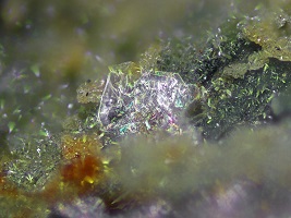
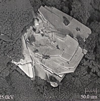
Specimen Size: 0.3 mm paper-thin plate of trogerite on pharmacosiderite
Field Collected: Bob Janules
Owner: Tom Mortimer
Catalog No.: u2089
Analysis: EDS analyzed, #BJ0076, J. Nizamoff, Aug, 2003.
No response was observed when this specimen was scanned with my scintilometer... indicating the total specimen uranium content was very low.
Notes: Very difficult to photograph this lusterous, reflective, plate. The choice is either a mirror bright reflection or vanishing transparent plate. The SEM image is very sharp, but limited to black and white.
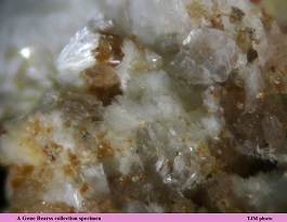
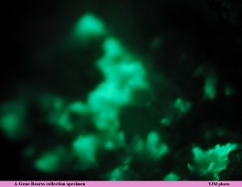
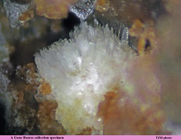
Confidence: 3
Chemistry: Ca2Be4(PO4)3(OH)3 · 5H2O
Locality: Palermo #1 Mine, N. Groton, NH
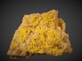
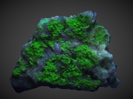
Specimen Size: 10 mm field of view. Normal and SW UV illumination. Zoom view 2 mm. (Tom Mortimer photos)
Field Collected: Acquired by Gene Bearss - 1992
Owner: Likely the Maine Mineral Museum - acquired Gene's mounted micro collection.
Analysis: None beyond visual observations noted below.
Notes: Uralolite is not listed in Bob Whitmore's book, The Pegmatite Mines Known as Palermo. Moraesite, Be2(PO4)(OH) · 4H2O, is listed. Uralolite, Ca2Be4(PO4)3(OH)3 · 5H2O, and moraesite are visually indestinguishable, however, uralolite is fluorescent while moraesite is not. Hydroxylherderite, another beryllium mineral, is also present on this specimen. These acicular crystals have fluorescent tips with non-fluorescent lower portion. Perhaps a zoned moreasite-uralolite? (but I am uncertain if this is structurally possible).
UV photos with the microscope require very long exposures.
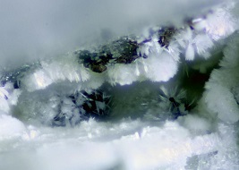
Confidence: 5
Locality: Turner Mine, Marlow, NH
Specimen Size: 1.3 mm field of view
Field Collected: Bob Wilken specimen & photo
Owner: Bob Wilken
Catalog No.: TUR02AS
Notes: An Al Falster of the MMGM lab EDS analysis indicated a mineral chemistry consistent with an uralolite identification. EDS cannot detect beryllium.
Locality: Turner Mine, Marlow, NH
Specimen Size: 4 mm field of view
Field Collected: Bob Wilken
Catalog No.: u2507
Notes: The uralolite is fluorescent green.
Locality: Turner Mine, Marlow, NH
Specimen Size: 1.5 mm field of view
Field Collected: Bob Wilken
Catalog No.: TBC
Notes: The uralolite is fluorescent green.
Confidence: 3
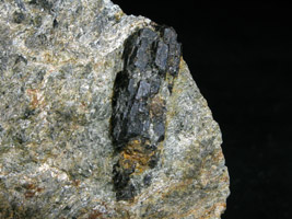
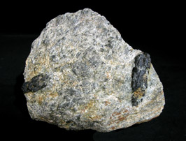
Chemistry: Ca0.75Na0.25Mg2.25Fe0.75Al6(BO3)3(Si6O18)(OH)3F
Locality: Tower Hill Rhodonite Locale, Hinsdale, NH
Specimen Size: 3 cm uvite crystal, largest on 9.5 cm specimen.
Field Collected - Owner: Tom Mortimer - May 5, 2005
Catalog No.: 1480
Analysis: Uvite-Dravite suggested by initial EDS analysis. Note in the analysis plot, the Mg content is much higher than the Fe content.
A second EDS analysis of this tourmaline with an instrument with good light element capability indicated a small sodium peak at 1.04 KeV, (not labeled on plot). Sodium is an "essential element" for dravite. End member dravite does not have calcium, which is an "essential element" for uvite. Both plots for this specimen show modest calcium .... and also iron, a component of schorl tourmaline. The plots do clearly show a magnesium dominant tourmaline. Both dravite and uvite satisfy this criteria, but uvite has a significant calcium content.
A third EDS analysis of this tourmaline, (3/2/16), including ZAF corrected element atomic percents, showed a Ca dominance over Na, (> 7X) indicating Uvite tourmaline.
Webmineral emperical formulae for Dravite & Uvite derived from analyses are given below for comparison with the APFU computed from this last EDS analysis, (adjusted for 3.4% boron, not detectable by the instrument, as a "best fit"):
Dravite: NaMg3Fe0.75Al5(BO3)3(Si6O18)(OH)
Uvite: Ca0.75Na0.25Mg2.25Fe0.75Al6(BO3)3(Si6O18)(OH)3F
#1480: Ca0.57Na0.14Mg1.9Fe0.55Al4.7(BO3)2.55(Si6O17)(OH)0
Notes: Uvite was included Janet Cares' New Hampshire species list published in vol. 65, no. 4 (Jul,Aug 1990) Rocks & Minerals, but I have been unable to locate a literature reference to substantiate this claim. Neither Phillip Morrill's New Hampshire Mines and Mineral Localities nor Meyers and Stewart's Part III Minerals and Mines make a reference to NH uvite.
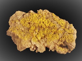
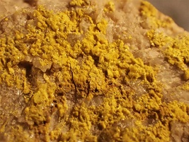
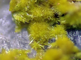
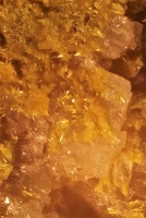
Confidence: 5
Chemistry: U6+(UO2)3(PO4)2(OH)6 · 4H2O.
Locality: Palermo #1 Mine, N. Groton, NH
Specimen Size: 1.6 x 1.4 cm specimen.
Field Collected Don Dallaire - 1983
Catalog No.: A Don Dallaire specimen and photos. (Micro crystal photo - Tom Mortimer)
Notes: [dd] "Sight id had it as uranophane since it was yellow and radioactive. It appeared to be a crust when I found it but with a better microscope than what I had at the time I saw balls of radiating crystals. I was also suspicious however since it fluoresced a bright green (uranophane has weak fluorescence if any at all). I brought it up to Al Falster [Maine Mineral Museum lab] on my monthly trip and he analyzed it. The EDS scan showed uranium and phosphorous and nothing else (no Na, Ca, Zn, Ba, Pb, Al, Sr, Fe, Cu, K, Mn, Mg or Nd). There were only 5 minerals that fit this. Four were quickly eliminated by other physical characteristics. The crystal is pseudohexagonal in outline but actually is orthorhombic. He tested the refractive indices and all 3 were greater than 1.7 (didn't have liquids for higher).
All this info fits the mineral vanmeersscheite: U6+(UO2)3(PO4)2(OH)6 · 4H2O."
A follow-up email from Don stated that the vanmeersscheite also has a dim green fluorescence in mid-wave and long-wave UV.
Locality: Chickering Mine, Walpole, NH
Confidence: 3
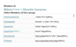
Chemistry: See group chemistry in second image.
Specimen Size: 10 mm field of view
Field Collected: Tom Mortimer
Catalog No.: u2298
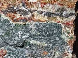
Notes: Dark green-black massive wyllieite group mineral. The wyllieite group species qingheiite is reported from the Chandlers Mill Mine in Newport, NH.
Prior to analysis, I thought this might be a compact form of frondelite, recently identified from the Chickering Mine.
Two polished grain EDS analyses computed a chemistry of: (normalized for 3 P)
Na1.4Fe0.84Mn0.19Mg0.75Ca0.16K0.12Al0.25P3O23
Na1.66Fe1.23Mn0.18Mg0.50Ca0.22K0.10Al0.26P3O36
This is a phosphate with dominant Na, Fe, and Mg cations. From the wyllieite group members, (tabulated below the specimen photo), it is seen that Ca. Fe, Mn, Mg can be distributed across multiple sites.
Confidence: 3
Chemistry: (Y,Ca,U4+,Fe2+)2(Ta,Nb)2O8
Locality: Ham-Weeks Mine, Wakefield, NH
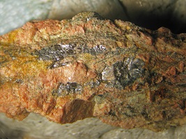
Specimen Size: 2 cm field of view. Resinous brown yttrotantalite-(Y) ? in red stained feldspar
Field Collected: Gene Bearss
Owner: Tom Mortimer
Catalog No.: u1498
Analysis: Yttrotantalite-(Y) suggested by EDS analysis . The initial consideration was for the samarskite-(Y) species. My samarskite group knowledgeable friend, Fred Davis, stated "I would be very hesitant to include a Ta>Nb mineral in the samarskite group." More analytic work is recommended for this specimen.
Notes: Yttrotantalite-(Y) has not previously been reported from New Hampshire.